Saturday 9 September 2017

CATARACTS - ALL YOU NEED TO KNOW

 SOURCE - WITH THE FULL ARTICLE

What is a cataract?


A cataract is an eye disease in which the clear lens of the eye becomes cloudy or opaque, causing a decrease in vision. Although the word cataract to describe this condition has been part of the English language since only the 15th century, the eye disease has been recognized and surgically treated since ancient times.

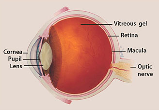
Eyeball Illustration - Cataracts
The lens is a portion of the eye that is normally clear. It focuses rays of light entering the eye onto the retina, the light-sensitive tissue at the back of the eye. In order to get a clear image onto the retina, the portions of the eye in front of the retina, including the lens, must be clear and transparent. The light striking the retina initiates a chemical reaction within the retina. The chemical reaction, in turn, initiates an electrical response which is carried to the brain through the optic nerve. The brain then interprets what the eye sees.

In a normal eye, light passes through the transparent lens to the retina. The lens must be clear for the retina to receive a sharp image. If the lens is cloudy from a cataract, the image striking the retina will be blurry or distorted and the vision will be blurry. The extent of the visual disturbance is dependent upon the degree of cloudiness of the lens.

Most cataracts are related to aging. Cataracts are very common in older people. By age 80, more than half of all Americans either have some degree of cataract or have already undergone cataract surgery in one or both eyes. By age 95, this percentage increases to almost 100%. A cataract can occur in either or both eyes. Individuals with a cataract in one eye usually go on to develop a cataract in the other eye as well. A cataract is not contagious and cannot spread from one eye to the other or from person to person. Cataracts do not cause the eye to tear abnormally. They are neither painful nor make the eye itchy or red.

Although vision can be restored in most people with cataracts, age-related cataracts are still the most common cause of blindness in the world, primarily because many third-world nations lack appropriate and available surgical services.
As life span increases in the developed world due to modern technology and new methods of treatment of acute and chronic disease, the incidence of age-related cataracts will continue to increase.

What are the different types of cataracts?


Cataracts can be classified by anatomical location within the lens, degree of clouding of the lens, or by the cause of the cataract.

The lens of the human eye is shaped and sized similar to an M&M candy. It has a front (anterior) part and a back (posterior) part. The central portion of the lens is called the lens nucleus, and the outer portion is called the lens capsule. Between the inner nucleus and the outer capsule is a portion of the lens called the cortex. Clouding of the lens can occur only in the nucleus, in which case the term "nuclear cataract" or "nuclear sclerosis" is used. If the clouding occurs in the lens cortex only, the cataract is termed a "cortical cataract." If the loss of clarity of the lens is primarily in or adjacent to the capsule, the term "subcapsular cataract" is used. The location of the clouding can also be defined as being anterior or posterior, central, or peripheral. Often the clouding of the lens may affect multiple portions of the lens. The most common type of cataract that is related to age is sometimes termed a "senile cataract." This type of cataract primarily involves the nucleus of the lens. Cataracts that develop in the posterior subcapsular area (in the rear region of the lens capsule) are more common in younger age groups.

Any degree of loss of the normal transparency of the lens is called a cataract. The cloudier the lens, the more advanced the degree of cataract. A cataract may be mild, moderate, or severe. It may be early or advanced. If the lens is totally opaque it is termed a "mature" cataract. Any cataract that is not opaque is therefore termed an "immature" cataract. Most mature cataracts are white in color.

What are risk factors for cataracts?


Advanced age is a significant risk factor for the development of cataracts. A family history for early development of cataracts, the presence of diabetestobacco use, and prolonged exposure to sunlight are also risk factors, as is trauma to the eye.

What are causes of cataracts?


The lens is made of mostly water and protein. The protein is arranged in a specific way that keeps the lens clear and allows light to pass through it to focus a clear image onto the retinal surface. As we age, some of the protein may clump together and start to cloud a small area of the lens. This is our understanding of the cause of an age-related cataract. 

Over time, the cataract may become denser or cloud more area of the lens, making it more difficult to see through. A cataract is not a growth or tumor.

There are many other causes of cataracts beyond advancing age. Whatever the cause, all cataracts are a result of similar changes to the protein of the lens, similarly resulting in visual blurring or visual loss.

Blunt or penetrating injury to the eye may result in a cataract, either immediately after the injury or some weeks to years afterward. A cataract following an injury may appear and then not increase in density (be stationary) or be progressive. Eye surgery for other conditions can also cause cataracts. Excessive exposure to ionizing radiation (X-ray), infrared radiation (as in glass blowers), or ultraviolet radiation may also cause cataracts.
Diabetes is associated with the development of cataracts. Inflammatory disease of the eye, such as iritis or uveitis, may cause or accelerate the development of cataract in the involved eye.

There are many genetic illnesses that are associated with the development of cataracts. These include myotonic dystrophy, galactosemia, homocystinuria, Wilson's disease and Down syndrome, plus many others. Congenital infections with herpes simplex, rubella,toxoplasmosissyphilis, and cytomegalic inclusion disease may also result in cataracts.

There are many medications which, when taken over a long period of time, can cause secondary cataracts. The most common of these are oral corticosteroids, such as prednisone, which currently are used for a wide variety of medical conditions.

The term "congenital cataract" is used when a baby is born with any clouding of the lens. This may be present in one or both eyes, be stationary or be progressive. Causes include genetic disorders or infectious or non-infectious intrauterine developmental disorders, both often associated with other physical abnormalities of the baby.
Atopic dermatitis, other diseases of the skin and mucous membranes, hypothyroidism, and hyperparathyroidism are associated with the early development of cataracts.

Patients who develop cataracts in both eyes at an early age often have family members who have also developed cataracts prematurely, implying a genetic cause, even in the absence of a recognized underlying disease.

What are the symptoms of cataracts?


You may not notice any symptoms with early cataracts. As a cataract becomes more advanced, decrease in clarity of vision, not fully correctable with glasses, is noticed. There is a loss of contrast sensitivity, so that shadows and color vision are less vivid. Disturbing glare may be noted as light is into the eye scattered by the cataract. Haloes may be observed around lights. Night vision will be diminished. In certain types of cataracts, double vision may be noted in the affected eye. Some patients note that they require frequent changes in their eyeglass or contact lens prescriptions and may be aware that their near vision is improving as their distance vision declines.
A cataract does not routinely cause discomfort or pain in the eye or alter the external appearance of the eye

What are the signs of cataracts?


Family members of a person affected by cataracts in both eyes may notice that he or she appears not to be seeing as well as previously. The eye will appear normal to the untrained observer, unless the cataract is mature and white. In that situation, the pupil of the eye, which normally appears black, will look grey or white to the observer.

The examining physician will find diminished visual acuity in the affected eye or eyes.

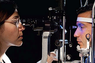
This visual loss is not fully corrected by a change in the glasses. The lens of the eye can be easily examined by an ophthalmologist and the changes in the lens characteristic of cataract can actually be seen using a slit lamp, which is an illuminating and magnifying device used to painlessly examine the structures within the front of the eye, including the lens.


What types of specialists treat cataracts?


Ophthalmologists are medical doctors who have specialized in the diagnosis and medical and surgical treatment of eye disease. Ophthalmologists both diagnose cataracts and surgically remove cataracts when indicated.


How do health-care professionals diagnose cataracts?


Cataracts are relatively simple to diagnose by an ophthalmologist or an optometrist during a routine eye examination. It is important, when making the diagnosis of cataract, to also examine the entire eye for evidence of any other eye disease which may be compromising the vision. In addition to taking a medical and ocular history and visual acuity test, the ophthalmologist will check eye movements and pupillary responses, measure the pressure inside the eyes and examine the both front and back of the eyes after the pupils have been dilated with drops.

What is the treatment for cataracts?


People with early cataract will find that changing their glasses, using sunglasses to decrease glare and having better lighting to read can significantly alleviate their symptoms. Magnifying lenses for close work and reading fine print may also be helpful.
Many cataracts are not bothersome, causing few symptoms. In that situation, no surgical treatment is necessary. However, the only true treatment for cataract is surgical removal of the cloudy lens. Surgery is suggested if the patient loses the ability to perform necessary activities of everyday life, such as driving, reading, or looking at computer or video screens, even with glasses, and there is the expectation that vision will improve as a result of the surgery.

Patients' responses to the presence of a cataract vary. A cataract in only one eye may be disturbing to a particular patient and may not cause significant symptoms in another patient.

Cataracts usually do not harm your eye, so you can have surgery when it is convenient for you and when the cataract interferes with your daily activities. Once you understand the benefits and risks of surgery, you can make an informed decision about whether cataract surgery is right for you. In most cases, delaying cataract surgery will not cause long-term damage to your eye or make the surgery more difficult.

If the eye has other diseases that have caused visual loss such as glaucomamacular degenerationdiabetic retinopathy, or optic nerve damage from glaucoma or other disease, cataract surgery may not improve the vision.
Occasionally, your doctor may recommend removal of a cataract if it prevents the diagnosis or treatment of another eye problem, such as macular degeneration or diabetic retinopathy.

If both eyes have cataracts and surgery is agreed upon, the surgery on the second eye is generally planned at least a week after the first eye. There is usually no harm in waiting a much longer period of time between the two eye operations.

Because the lens of the eye is necessary to accurately focus light onto the retinal surface and removal of the cataract involves removal of the lens, modern cataract surgery combines removal of the lens and placement of a new artificial lens into the eye.

Measurements for the size, shape, and power of this lens will be taken prior to the surgery so that the specific lens can be available for implantation at the time of surgery.

More than 3 million cataract surgeries are performed annually in the United States. Cataract surgery is extremely safe and effective, improving vision in the vast majority of patients.

What are risks of the different types of cataract surgery? 


How long is the recovery after cataract surgery?


Cataract surgery is usually performed as an outpatient procedure under local anesthesia. Some sedation is ordinarily given intravenously just before the beginning of the surgery, which usually takes under a half-hour.


Most cataract surgery today is done through a small incision by phaco-emulsification or by other means through a slightly larger incision. In more than 95% of cases, a new lens, known as a lens implant or intraocular lens is inserted at the same time as the cataract removal. You will not feel the new lens within the eye. Most patients need to limit their activities for only a few days and recovery time is brief.


Although modern techniques have made cataract surgery quite safe, complications can occur with any surgical procedure, including cataract extraction. These include hemorrhage, infection, loss of a portion of the cataract into the eye, displacement of the intraocular lens, glaucoma, and retinal detachment. Fortunately, all these complications are rare and usually can be managed. Blindness is a rare complication of cataract surgery.

Modern cataract surgery involves leaving a portion of the lens capsule within the eye to support the intraocular lens. This capsule may become cloudy at a later time, necessitating opening of the capsule through the use of a laser. This outpatient procedure is called a YAG laser capsulotomy. It is painless and rarely results in increased eye pressure or other eye problems

What are complications of cataracts?


Occasionally, a very dense cataract of long-standing duration may enlarge in size and interfere with fluid drainage within the eye. In addition, a far advanced cataract may leak protein into the eye, causing inflammation of the eye. Your doctor will advise you of these possibilities and may recommend surgery to avoid these complications even if the decrease in vision is not bothering you.
The presence of a cataract may make evaluation of diseases of the retina more difficult, since the physician must look through the cataract to examine the retina.


What is the prognosis of cataracts?


The rate of progression of cataracts is usually predictable and surgery is successful in restoring vision in a large majority of cases. If other diseases are present within the eye, the degree of visual improvement will be limited by the other disease process. Ophthalmologists can usually determine this in advance.


Is it possible to prevent cataracts?


Everyone, if they live long enough, will develop cataracts. There is no scientifically proven method that prevents the inevitable. Progression of cataracts can be slowed by avoiding large amounts of ultraviolet light, not smoking, avoiding trauma to either eye, and following a healthy diet. Wearing UV-protection sunglasses when exposed to sunlight can be helpful.

Where can people get more information on cataracts?

Cataract Surgery: A Patient's Guide to Cataract Treatment by Julius Shulman, MD

REFERENCES:

Asbell, P.A., I. Dualan, J. Mindel, et al. "Age-related cataract." Lancet 365 (2005): 599.

Cataracts InfoCenter. <http://www.cataractsinfocenter.com/>.

Congdon, N., J.R. Vingerling, B.E. Klein, et al. "Prevalence of cataract and pseudophakia/aphakia among adults in the United States." Arch Ophthalmol 122 (2004): 487.

Lai, F.H., J.Y. Lok, P.P. Chow, and A.L. Young. "Clinical outcomes of cataract surgery in very elderly adults." J Am Geriatr Soc 62 (2014): 165.

Mares, J.A., R. Voland, R. Adler, et al. "Healthy diets and the subsequent prevalence of nuclear cataract in women." Arch Ophthalmol 128 (2010): 738.

United States. National Eye Institute. National Institutes of Health. "Facts About Cataract." Feb. 2010. <http://www.nei.nih.gov/health/cataract/cataract_facts.asp>.
Last Editorial Review: 8/12/2016

DIABETIC RETINOPATY

 ORIGINAL SOURCE


Diabetic retinopathy is a condition that occurs in people who have diabetes. It causes progressive damage to the retina, the light-sensitive lining at the back of the eye. Diabetic retinopathy is a serious sight-threatening complication of diabetes.

Diabetes interferes with the body's ability to use and store sugar (glucose). The disease is characterized by too much sugar in the blood, which can cause damage throughout the body, including the eyes.

Over time, diabetes damages the blood vessels in the retina. Diabetic retinopathy occurs when these tiny blood vessels leak blood and other fluids. This causes the retinal tissue to swell, resulting in cloudy or blurred vision. The condition usually affects both eyes. The longer a person has diabetes, the more likely they will develop diabetic retinopathy. If left untreated, diabetic retinopathy can cause blindness.

Symptoms of diabetic retinopathy include:
  • Seeing spots or floaters
  • Blurred vision
  • Having a dark or empty spot in the center of your vision
  • Difficulty seeing well at night
When people with diabetes experience long periods of high blood sugar, fluid can accumulate in the lens inside the eye that controls focusing. This changes the curvature of the lens, leading to blurred vision. However, once blood sugar levels are controlled, blurred distance vision will improve. Patients with diabetes who can better control their blood sugar levels will slow the onset and progression of diabetic retinopathy.

Often the early stages of diabetic retinopathy have no visual symptoms. That is why the American Optometric Association recommends that everyone with diabetes have a comprehensive dilated eye examination once a year. Early detection and treatment can limit the potential for significant vision loss from diabetic retinopathy.

Treatment of diabetic retinopathy varies depending on the extent of the disease. People with diabetic retinopathy may need laser surgery to seal leaking blood vessels or to discourage other blood vessels from leaking. Your optometrist might need to inject medications into the eye to decrease inflammation or stop the formation of new blood vessels. People with advanced cases of diabetic retinopathy might need a surgical procedure to remove and replace the gel-like fluid in the back of the eye, called the vitreous. Surgery may also be needed to repair a retinal detachment. This is a separation of the light-receiving lining in the back of the eye.

If you are diabetic, you can help prevent or slow the development of diabetic retinopathy by:
  • Taking your prescribed medication
  • Sticking to your diet
  • Exercising regularly
  • Controlling high blood pressure
  • Avoiding alcohol and smoking

What causes diabetic retinopathy?

Non-proliferative diabetic retinopathy (NPDR) is the early state of the disease in which symptoms will be mild or non-existent. In NPDR, the blood vessels in the retina are weakened causing tiny bulges called microanuerysms to protrude from their walls.

Proliferative diabetic retinopathy (PDR) is the more advanced form of the disease. At this stage, new fragile blood vessels can begin to grow in the retina and into the vitreous, the gel-like fluid that fills the back of the eye. The new blood vessel may leak blood into the vitreous, clouding vision.
Diabetic retinopathy results from the damage diabetes causes to the small blood vessels located in the retina. These damaged blood vessels can cause vision loss:
  • Fluid can leak into the macula, the area of the retina responsible for clear central vision. Although small, the macula is the part of the retina that allows us to see colors and fine detail. The fluid causes the macula to swell, resulting in blurred vision.
  • In an attempt to improve blood circulation in the retina, new blood vessels may form on its surface. These fragile, abnormal blood vessels can leak blood into the back of the eye and block vision.
Diabetic retinopathy is classified into two types:
  1. Non-proliferative diabetic retinopathy (NPDR) is the early stage of the disease in which symptoms will be mild or nonexistent. In NPDR, the blood vessels in the retina are weakened. Tiny bulges in the blood vessels, called microaneurysms, may leak fluid into the retina. This leakage may lead to swelling of the macula.
  2. Proliferative diabetic retinopathy (PDR) is the more advanced form of the disease. At this stage, circulation problems deprive the retina of oxygen. As a result new, fragile blood vessels can begin to grow in the retina and into the vitreous, the gel-like fluid that fills the back of the eye. The new blood vessels may leak blood into the vitreous, clouding vision.
Other complications of PDR include detachment of the retina due to scar tissue formation and the development of glaucoma. Glaucoma is an eye disease in which there is progressive damage to the optic nerve. In PDR, new blood vessels grow into the area of the eye that drains fluid from the eye. This greatly raises the eye pressure, which damages the optic nerve. If left untreated, PDR can cause severe vision loss and even blindness.

Risk factors for diabetic retinopathy include:
  • Diabetes. People with type 1 or type 2 diabetes are at risk for developing diabetic retinopathy. The longer a person has diabetes, the more likely he or she is to develop diabetic retinopathy, particularly if the diabetes is poorly controlled.
  • Race. Hispanics and African Americans are at greater risk for developing diabetic retinopathy.
  • Medical conditions. People with other medical conditions, such as high blood pressure and high cholesterol, are at greater risk.
  • Pregnancy. Pregnant women face a higher risk for developing diabetes and diabetic retinopathy. If a woman develops gestational diabetes, she has a higher risk of developing diabetes as she ages.

How is diabetic retinopathy diagnosed?

Comprehensive eye exam
Diabetic retinopathy can be diagnosed through a comprehensive eye examination. Testing, with emphasis on evaluating the retina and macula, may include:
  • Patient history to determine vision difficulties, presence of diabetes, and other general health concerns that may be affecting vision
  • Visual acuity measurements to determine how much central vision has been affected
  • Refraction to determine if a new eyeglass prescription is needed
  • Evaluation of the ocular structures, including the evaluation of the retinathrough a dilated pupil
  • Measurement of the pressure within the eye
Supplemental testing may include:
  • Retinal photography or tomography to document current status of the retina
  • Fluorescein angiography to evaluate abnormal blood vessel growth
How is diabetic retinopathy treated?
Laser treatment (photocoagulation) is used to stop the leakage of blood and fluid into the retina. A laser beam of light can be used to create small burns in areas of the retina with abnormal blood vessels to try to seal the leaks.
Treatment for diabetic retinopathy depends on the stage of the disease. The goal of any treatment is to slow or stop the progression of the disease.

In the early stages of non-proliferative diabetic retinopathy, regular monitoring may be the only treatment. Following your doctor's advice for diet and exercise and controlling blood sugar levels can help control the progression of the disease.

If the disease advances, the blood vessels can leak blood and fluid into the retina, leading to macular edema. Laser treatment (photocoagulation) can stop this leakage. A laser beam of light creates small burns in areas of the retina with abnormal blood vessels to try to seal the leaks.

Widespread blood vessel growth in the retina, which occurs in proliferative diabetic retinopathy, can be treated by creating a pattern of scattered laser burns across the retina. This causes abnormal blood vessels to shrink and disappear. With this procedure, some side vision may be lost in order to safeguard central vision.

GLAUCOMA

 SOURCE


Facts About Glaucoma


This information was developed by the National Eye Institute to help patients and their families search for general information about glaucoma. An eye care professional who has examined the patient’s eyes and is familiar with his or her medical history is the best person to answer specific questions.

Glaucoma Defined

What is Glaucoma?

Glaucoma is a group of diseases that damage the eye’s optic nerve and can result in vision loss and blindness. However, with early detection and treatment, you can often protect your eyes against serious vision loss.
The optic nerve
The optic nerve
The optic nerve is a bundle of more than 1 million nerve fibers. It connects the retina to the brain. (See diagram above.) The retina is the light-sensitive tissue at the back of the eye. A healthy optic nerve is necessary for good vision.

How does the optic nerve get damaged by open-angle glaucoma?

Several large studies have shown that eye pressure is a major risk factor for optic nerve damage. In the front of the eye is a space called the anterior chamber. A clear fluid flows continuously in and out of the chamber and nourishes nearby tissues. The fluid leaves the chamber at the open angle where the cornea and iris meet. (See diagram below.) When the fluid reaches the angle, it flows through a spongy meshwork, like a drain, and leaves the eye.
In open-angle glaucoma, even though the drainage angle is “open”, the fluid passes too slowly through the meshwork drain. Since the fluid builds up, the pressure inside the eye rises to a level that may damage the optic nerve. When the optic nerve is damaged from increased pressure, open-angle glaucoma-and vision loss—may result. That’s why controlling pressure inside the eye is important.
Another risk factor for optic nerve damage relates to blood pressure. Thus, it is important to also make sure that your blood pressure is at a proper level for your body by working with your medical doctor.
Fluid Pathway
Fluid pathway is shown in teal.

Can I develop glaucoma if I have increased eye pressure?

Not necessarily. Not every person with increased eye pressure will develop glaucoma. Some people can tolerate higher levels of eye pressure better than others. Also, a certain level of eye pressure may be high for one person but normal for another.
Whether you develop glaucoma depends on the level of pressure your optic nerve can tolerate without being damaged. This level is different for each person. That’s why a comprehensive dilated eye exam is very important. It can help your eye care professional determine what level of eye pressure is normal for you.

Can I develop glaucoma without an increase in my eye pressure?

Yes. Glaucoma can develop without increased eye pressure. This form of glaucoma is called low-tension or normal-tension glaucoma. It is a type of open-angle glaucoma.

Who is at risk for open-angle glaucoma?

Anyone can develop glaucoma. Some people, listed below, are at higher risk than others:
  • African Americans over age 40
  • Everyone over age 60, especially Mexican Americans
  • People with a family history of glaucoma
A comprehensive dilated eye exam can reveal more risk factors, such as high eye pressure, thinness of the cornea, and abnormal optic nerve anatomy. In some people with certain combinations of these high-risk factors, medicines in the form of eyedrops reduce the risk of developing glaucoma by about half.

Glaucoma Symptoms

At first, open-angle glaucoma has no symptoms. It causes no pain. Vision stays normal. Glaucoma can develop in one or both eyes.
Without treatment, people with glaucoma will slowly lose their peripheral (side) vision. As glaucoma remains untreated, people may miss objects to the side and out of the corner of their eye. They seem to be looking through a tunnel. Over time, straight-ahead (central) vision may decrease until no vision remains.
Normal Vision
Normal Vision.
Glaucoma Vision
The same scene as viewed by a person with glaucoma.
Glaucoma Detected

How is glaucoma detected?

Glaucoma is detected through a comprehensive dilated eye exam that includes the following:
Visual acuity test. This eye chart test measures how well you see at various distances.
Visual field test. This test measures your peripheral (side vision). It helps your eye care professional tell if you have lost peripheral vision, a sign of glaucoma.
Dilated eye exam. In this exam, drops are placed in your eyes to widen, or dilate, the pupils. Your eye care professional uses a special magnifying lens to examine your retina and optic nerve for signs of damage and other eye problems. After the exam, your close-up vision may remain blurred for several hours.
Tonometry is the measurement of pressure inside the eye by using an instrument called a tonometer. Numbing drops may be applied to your eye for this test. A tonometer measures pressure inside the eye to detect glaucoma.
Pachymetry is the measurement of the thickness of your cornea. Your eye care professional applies a numbing drop to your eye and uses an ultrasonic wave instrument to measure the thickness of your cornea.

Can glaucoma be cured?

No. There is no cure for glaucoma. Vision lost from the disease cannot be restored.

Glaucoma Treatments

Immediate treatment for early-stage, open-angle glaucoma can delay progression of the disease. That’s why early diagnosis is very important.
Glaucoma treatments include medicines, laser trabeculoplasty, conventional surgery, or a combination of any of these. While these treatments may save remaining vision, they do not improve sight already lost from glaucoma.
Medicines. Medicines, in the form of eyedrops or pills, are the most common early treatment for glaucoma. Taken regularly, these eyedrops lower eye pressure. Some medicines cause the eye to make less fluid. Others lower pressure by helping fluid drain from the eye.
Before you begin glaucoma treatment, tell your eye care professional about other medicines and supplements that you are taking. Sometimes the drops can interfere with the way other medicines work.
Glaucoma medicines need to be taken regularly as directed by your eye care professional. Most people have no problems. However, some medicines can cause headaches or other side effects. For example, drops may cause stinging, burning, and redness in the eyes.
Many medicines are available to treat glaucoma. If you have problems with one medicine, tell your eye care professional. Treatment with a different dose or a new medicine may be possible.
Because glaucoma often has no symptoms, people may be tempted to stop taking, or may forget to take, their medicine. You need to use the drops or pills as long as they help control your eye pressure. Regular use is very important.
Tonometer that measures pressure.
A tonometer measures pressure inside the eye to detect glaucoma.
Make sure your eye care professional shows you how to put the drops into your eye. For tips on using your glaucoma eyedrops, see the inside back cover of this booklet.
Laser trabeculoplasty. Laser trabeculoplasty helps fluid drain out of the eye. Your doctor may suggest this step at any time. In many cases, you will need to keep taking glaucoma medicines after this procedure.
Laser trabeculoplasty is performed in your doctor’s office or eye clinic. Before the surgery, numbing drops are applied to your eye. As you sit facing the laser machine, your doctor holds a special lens to your eye. A high-intensity beam of light is aimed through the lens and reflected onto the meshwork inside your eye. You may see flashes of bright green or red light. The laser makes several evenly spaced burns that stretch the drainage holes in the meshwork. This allows the fluid to drain better.
Like any surgery, laser surgery can cause side effects, such as inflammation. Your doctor may give you some drops to take home for any soreness or inflammation inside the eye. You will need to make several follow-up visits to have your eye pressure and eye monitored.
If you have glaucoma in both eyes, usually only one eye will be treated at a time. Laser treatments for each eye will be scheduled several days to several weeks apart.
Studies show that laser surgery can be very good at reducing the pressure in some patients. However, its effects can wear off over time. Your doctor may suggest further treatment.
Conventional surgery. Conventional surgery makes a new opening for the fluid to leave the eye. (See diagram on the next page.) Your doctor may suggest this treatment at any time. Conventional surgery often is done after medicines and laser surgery have failed to control pressure.
Conventional surgery, called trabeculectomy, is performed in an operating room. Before the surgery, you are given medicine to help you relax. Your doctor makes small injections around the eye to numb it. A small piece of tissue is removed to create a new channel for the fluid to drain from the eye. This fluid will drain between the eye tissue layers and create a blister-like “filtration bleb.”
For several weeks after the surgery, you must put drops in the eye to fight infection and inflammation. These drops will be different from those you may have been using before surgery.
Conventional surgery is performed on one eye at a time. Usually the operations are four to six weeks apart.
Conventional surgery is about 60 to 80 percent effective at lowering eye pressure. If the new drainage opening narrows, a second operation may be needed. Conventional surgery works best if you have not had previous eye surgery, such as a cataract operation.
Sometimes after conventional surgery, your vision may not be as good as it was before conventional surgery. Conventional surgery can cause side effects, including cataract, problems with the cornea, inflammation, infection inside the eye, or low eye pressure problems. If you have any of these problems, tell your doctor so a treatment plan can be developed.

What are some other forms of glaucoma and how are they treated?

Open-angle glaucoma is the most common form. Some people have other types of the disease.
In low-tension or normal-tension glaucoma, optic nerve damage and narrowed side vision occur in people with normal eye pressure. Lowering eye pressure at least 30 percent through medicines slows the disease in some people. Glaucoma may worsen in others despite low pressures.
A comprehensive medical history is important to identify other potential risk factors, such as low blood pressure, that contribute to low-tension glaucoma. If no risk factors are identified, the treatment options for low-tension glaucoma are the same as for open-angle glaucoma.
In angle-closure glaucoma, the fluid at the front of the eye cannot drain through the angle and leave the eye. The angle gets blocked by part of the iris. People with this type of glaucoma may have a sudden increase in eye pressure. Symptoms include severe pain and nausea, as well as redness of the eye and blurred vision. If you have these symptoms, you need to seek treatment immediately. This is a medical emergency. If your doctor is unavailable, go to the nearest hospital or clinic. Without treatment to restore the flow of fluid, the eye can become blind. Usually, prompt laser surgery and medicines can clear the blockage, lower eye pressure, and protect vision.
In congenital glaucoma, children are born with a defect in the angle of the eye that slows the normal drainage of fluid. These children usually have obvious symptoms, such as cloudy eyes, sensitivity to light, and excessive tearing. Conventional surgery typically is the suggested treatment, because medicines are not effective and can cause more serious side effects in infants and be difficult to administer. Surgery is safe and effective. If surgery is done promptly, these children usually have an excellent chance of having good vision.
Conventional surgery.
Conventional surgery makes a new opening for the fluid to leave the eye.
Secondary glaucomas can develop as complications of other medical conditions. For example, a severe form of glaucoma is called neovascular glaucoma, and can be a result from poorly controlled diabetes or high blood pressure. Other types of glaucoma sometimes occur with cataract, certain eye tumors, or when the eye is inflamed or irritated by a condition called uveitis. Sometimes glaucoma develops after other eye surgeries or serious eye injuries. Steroid drugs used to treat eye inflammations and other diseases can trigger glaucoma in some people. There are two eye conditions known to cause secondary forms of glaucoma.
Pigmentary glaucoma occurs when pigment from the iris sheds off and blocks the meshwork, slowing fluid drainage.
Pseudoexfoliation glaucoma occurs when extra material is produced and shed off internal eye structures and blocks the meshwork, again slowing fluid drainage.
Depending on the cause of these secondary glaucomas, treatment includes medicines, laser surgery, or conventional or other glaucoma surgery.

What research is being done?

Through studies in the laboratory and with patients, NEI is seeking better ways to detect, treat, and prevent vision loss in people with glaucoma. For example, researchers have discovered genes that could help explain how glaucoma damages the eye.
NEI also is supporting studies to learn more about who is likely to get glaucoma, when to treat people who have increased eye pressure, and which treatment to use first.

What You Can Do

If you are being treated for glaucoma, be sure to take your glaucoma medicine every day. See your eye care professional regularly.
You also can help protect the vision of family members and friends who may be at high risk for glaucoma-African Americans over age 40; everyone over age 60, especially Mexican Americans; and people with a family history of the disease. Encourage them to have a comprehensive dilated eye exam at least once every two years. Remember that lowering eye pressure in the early stages of glaucoma slows progression of the disease and helps save vision.
Medicare covers an annual comprehensive dilated eye exam for some people at high risk for glaucoma. These people include those with diabetes, those with a family history of glaucoma, and African Americans age 50 and older.

What should I ask my eye care professional?

You can protect yourself against vision loss by working in partnership with your eye care professional. Ask questions and get the information you need to take care of yourself and your family.

What are some questions to ask?

About my eye disease or disorder…
  • What is my diagnosis?
  • What caused my condition?
  • Can my condition be treated?
  • How will this condition affect my vision now and in the future?
  • Should I watch for any particular symptoms and notify you if they occur?
  • Should I make any lifestyle changes?
About my treatment…
  • What is the treatment for my condition?
  • When will the treatment start and how long will it last?
  • What are the benefits of this treatment and how successful is it?
  • What are the risks and side effects associated with this treatment?
  • Are there foods, medicines, or activities I should avoid while I’m on this treatment?
  • If my treatment includes taking medicine, what should I do if I miss a dose?
  • Are other treatments available?
About my tests…
  • What kinds of tests will I have?
  • What can I expect to find out from these tests?
  • When will I know the results?
  • Do I have to do anything special to prepare for any of the tests?
  • Do these tests have any side effects or risks?
  • Will I need more tests later?
Other suggestions
  • If you don’t understand your eye care professional’s responses, ask questions until you do understand.
  • Take notes or get a friend or family member to take notes for you. Or, bring a tape recorder to help you remember the discussion.
  • Ask your eye care professional to write down his or her instructions to you.
  • Ask your eye care professional for printed material about your condition.
  • If you still have trouble understanding your eye care professional’s answers, ask where you can go for more information.
  • Other members of your healthcare team, such as nurses and pharmacists, can be good sources of information. Talk to them, too.
Today, patients take an active role in their health care. Be an active patient about your eye care.

Loss of Vision

If you have lost some sight from glaucoma, ask your eye care professional about low vision services and devices that may help you make the most of your remaining vision. Ask for a referral to a specialist in low vision. Many community organizations and agencies offer information about low vision counseling, training, and other special services for people with visual impairments.

How should I use my glaucoma eyedrops?

If eyedrops have been prescribed for treating your glaucoma, you need to use them properly, as instructed by your eye care professional. Proper use of your glaucoma medication can improve the medicine’s effectiveness and reduce your risk of side effects.
To properly apply your eyedrops, follow these steps:
  • Wash your hands.
  • Hold the bottle upside down.
  • Tilt your head back.
  • Hold the bottle in one hand and place it as close as possible to the eye.
  • With the other hand, pull down your lower eyelid. This forms a pocket.
  • Place the prescribed number of drops into the lower eyelid pocket. If you are using more than one eyedrop, be sure to wait at least 5 minutes before applying the second eyedrop.
  • Close your eye OR press the lower lid lightly with your finger for at least 1 minute. Either of these steps keeps the drops in the eye and helps prevent the drops from draining into the tear duct, which can increase your risk of side effects.

The National Eye Institute (NEI) is part of the National Institutes of Health (NIH) and is the Federal government’s lead agency for vision research that leads to sight-saving treatments and plays a key role in reducing visual impairment and blindness.