Tuesday 29 August 2017

PREVENTION OF BLINDNESS - MANAGEMENT AND SCIENCE UNIVERSITY EYE CENTRE

 MSU EYE CENTRE

Note : See Video on the right column

Blindness is one of the major issues confronting public health the world over. Studies reveal that the rate of worldwide blindness and visual impairment by 2020 is about 285 million and blindness cause by a single eye disease such as glaucoma will affect about 60 million people. In Malaysia, it is estimated that the overall prevalence of blindness and visual impairment is up to one (1) million people are suffering from blindness or vision related issues.


The Malaysian Eye disease registry 2012 data stated that more than 200,000 Malaysians suffer from blindness and the figure will increase in the coming years due to poor access and very low awareness to comprehensive eye examination among the population. A recent study conducted in State of Selangor shown the prevalence of visual impairment and blindness was up to 21% mainly caused by uncorrected refractive errors, cataract and diabetic retinopathy.

Parallel to its effort to seeing community wellness being given special importance, MSU through its Vision and Eye studies is committed to deal with blindness issue towards the creation of a better society free from such health challenges. The MSU Prevention of Blindness Project (MSU POB Project) was initiated to deal with awareness, promote vision screening for an early detection of eye and vision problems. The project was launched in 2012 with the University allocating RM300,000 to ensure the success of the undertaking.

This three (3) year project divided into two (2) phases with phase 1 involving the screening program covering children’s vision care, the urban poor and indigenous people, orphanages, the elderly and the industrial sectors. Phase 2, encompasses clinical intervention including cataract screening and surgery, training and research activities as well as public health services provided to those in need.

The MSU-POB Project is led by Associate Professor Dr Mohd Zaki Awg Isa assisted by three other researchers, Madam Fairuz Mohd Nordin and Rajasegar Anamalley and Madam Azarina Abdullah. The focus now (Phase 2) is more towards enhancing implementation of translational research by empowering community. In 2016, two niche areas of research were identified involving school children and ‘orang asli’. The activities involved are mass children vision screening program, vision research for ‘orang asli’ school children and initiating a special vision project in collaboration with Sekolah Kebangsaan U3 Subang Jaya, training the teachers for early detection of eye and vision problems in school and providing free spectacles and vision aids to the poor were also among the activities held.

The project was successful at engaging all parties involved this include manufactures, suppliers, NGO, Industries and local community through continuous eye health screening, campaigns, donation, participation and support to promote awareness in preventing blindness. The POB program also provides knowledge transfer to the community by providing avenues to the undergraduate and post graduate students to enhance their soft skills and competencies.

Likewise, the lecturers also have opportunities to conduct research projects in the prevention of blindness programs which later will benefits the communities at large.

Monday 28 August 2017

Preventive care can help you keep your eyes healthy and avoid common causes of blindness

ORIGINAL SOURCE : EVERYDAY HEALTH

WHO PREVENTION OF BLINDNESS - VISUAL IMPAIRMENT

 SOURCE
Excerpt

The most recent WHO estimates on the global magnitude and causes of visual impairments confirm a major opportunity for change in the lives of millions of people: 80% of all causes of visual impairment are preventable or curable. WHO estimates that in 2010 there were 285 million people visually impaired,
of which 39 million were blind. If just the two major causes of visual impairment were considered priorities and control measures were implemented consistently across the world, by providing refractive services and offering cataract surgery to the people in need, two thirds of the visually impaired people could recover good sight. This scenario appears to be fairly easy to realize, but for multiple reasons both the aforementioned eye diseases remain major items on the unfinished agenda of public eye care

Dr Oleg Chestnov
Assistant Director-General
Noncommunicable Diseases and Mental Health
World Health Organization

 DOWNLOAD WHO REPORT


Excerpt

Elimination of avoidable blindness depends on progress in other global health and development agendas, such as the development of comprehensive health systems, human resources for health development, improvements in the area of maternal, child and reproductive health, and the provision of safe drinking-water and basic sanitation. Eye health should be included in broader noncommunicable and communicable disease frameworks, as well as those addressing ageing populations. The proven risk factors for some causes of blindness (e.g. diabetes mellitus, smoking, premature birth, rubella and vitamin A deficiency) need to be continuously addressed through multisectoral interventions.

 DOWNLOAD WHO ANNEX 2


Thursday 24 August 2017

BINOCULAR VISION AMONG CHILDREN

 CHILDREN SPECIAL NEEDS

This web site was created to raise public awareness of treatment options for binocular vision impairments, such as amblyopia ("lazy eye") and strabismus (wandering eye, wall-eyes, crossed-eyes Crossed Eyes, Wandering Eye, Deviating Eyes, Esotropia, Exotropia, Esophoria, Exophoria, Hyperphoria, Hyperopia, Astigmatism, Alternating Esotropia), Convergence Insufficiency, Double Vision, Diplopia, Nystagmus, Loss of Depth Perception, Poor Binocular Vision, Accommodative Problems, Tracking Problems, Loss of Depth Perception, Loss of stereoscopic vision and/or binocular fusion, Learning Related Vision Problems.


What is Binocular Vision?


BINOCULAR: Of or involving both eyes at once.

BINOCULAR VISION: Vision wherein both eyes aim simultaneously at the same visual target; vision wherein both eyes work together -- simultaneously, equally and accurately -- as a coordinated team.

Healthy binocular vision produces important visual perceptual skills which are part of normal human vision: binocular depth perception and stereopsis.

STEREOPSIS: (stereopsis or stereoscopic vision) vision wherein two separate images from two eyes are successfully combined into one image in the brain.

Stereopsis is an aspect of "normal" healthy vision. Here's how it works. First, both eyes must be accurately aimed at the same target (that's binocular vision, but it's not yet stereoscopic vision!) Then, because the two eyes are located in different positions, each takes in a unique view from its own perspective. The two separate images are sent on to the brain for processing. When the two images arrive simultaneously in the back of the brain they are united into one picture. The combined picture appears three-dimensional (3-D) because it has the added depth dimension. That's stereo vision. Stereo vision gives you depth perception. For an illustrated definition, see the Stereo Vision page.

What is Binocular Vision Impairment?

A binocular vision impairment is any visual condition wherein binocular visual skills are inadequately developed. A comprehensive eye examination should include the testing of these important binocular visual skills:
Tracking: the ability to move the eyes across a sheet of paper
Fusion: the ability to use both eyes together at the same time
Stereopis: binocular depth perception
Convergence: the ability of the eyes to move and work as a team
Visual Motor Integration: the ability to transform images from a vertical to a horizontal plane

Binocular vision impairments often result in partial or total loss of stereoscopic vision and binocular depth perception. Conditions where the eye is obviously turned or crossed are commonly referred to with terms like "cross-eyed", "crossed-eyes", wall-eyes" or "wandering eyes". These binocular vision impairments are easily detected by others as all the observer needs to do is notice that both eyes do not aim in the same direction at all times. Some medical terms for these deviations or misalignments of the eye are strabismus, esotropia, exotropia, congenital strabismus, hereditary strabismus, pediatric strabismus, adult strabismus, accommodative esotropia, alternating esotropia, congenital esotropia, accommodative esotropia, esophoria, exophoria, exotropia, exotropic, esotropic, hyperphoria, hypophoria, hyperphoria, hypertropia. Other medical conditions which can contribute to poor binocular vision are anesmetropia, astigmatism, nearsightness, myopia, farsightedness, hyperopia, nystagmus, cerebral palsy, cataracts.

PLEASE NOTE: Some binocular vision impairments are not easily detected by parents, teacher or others because the turning or straying of the eye(s) is NOT obvious or consistent. Some eye turns are intermittent (they come and go) and/or they are not easily noticed by the untrained observer. A binocular vision problem is indicated, even when an eye turn is only occasionally visible. That's why early examination is so important.

Has your child been given different prescriptions for his or her two eyes? It is important to note that binocular vision is often impaired when the vision in the two eyes is significantly different or if one eye is higher than the other. The wearing of prescription glasses ONLY will rarely correct poor binocular vision.

Binocular vision impairments are more common than you may think. Just one type of binocular impairment, amblyopia ("lazy eye"), affects approximately 3% of the population. At least 12% of the population has some type of problem with binocular vision. Plenty of people are seeing the world with only one eye. They are monocular (one-eyed), not binocular (two-eyed). (Remember the monocles that people used to wear in the nineteenth century?)

Many monocular people can be rehabilitated with the help of Vision Therapy. They can become binocular and gain depth perception! children-special-needs.org seeks to call attention to binocular vision impairments and to educate parents and the public about treatment options. Many parents of children with these visual defects are not informed of all treatment options and are not gaining information about and access to Vision Therapy, a treatment option that offers a much higher success rate than eye muscle surgery.

What is Vision Therapy?

VISION THERAPY: (also known as vision training, visual training, visual therapy, eye training, behavioral optometry or orthoptic therapy):

Vision therapy can be described as physical therapy for the visual system which includes the brain and eyes. Through a series of progressive therapeutic procedures (eye exercises), patients develop or recover normal visual skills. Vision therapy is remarkably successful in rehabilitating all types of binocular vision impairments including amblyopia (lazy eye), strabismus, esotropia, exotropia, hyperphoria, or loss of binocular fusion due to hyperopia (farsightedness), myopia (nearsightedness) or astigmatism in one eye. In regards to the development or recovery of binocular vision, Vision Therapy is much more successful than surgery or glasses alone.

Who Can Benefit From Vision Therapy?

Patients of all ages can benefit from Vision Therapy. The nature of the therapy program varies with the condition treated. For example, a three year-old child with amblyopia, or "lazy eye", might have the better eye patched for a short period of time. An eight year old with strabismus "crossed eye", may require therapy for a period of a year to to gain BOTH cosmetic and visual benefits (the two eyes will appear straight AND will be used as a binocular team for normal vision). A 30 year-old computer programmer may require three to six months to solve a visual problem causing significant eye strain. A 30 year-old with strabismus who had two or more unsuccessful surgeries as a child might require two or more years of therapy. For information on patients with dyslexia, attention deficit disorder, and other conditions related to visual perception and information processing, see add-adhd.org.

Vision therapy can improve visual skills such as stereopsis, binocular coordination, binocular fusion, eye teaming skills, convergence, visual acuity, focusing skills, stereoscopic vision, depth perception, eye tracking, fixation skills, visual form discrimination, visual memory, hyperopia, and visual motor integration (balance, body coordination, hand-eye coordination).

Is Vision Therapy New?

Although Vision Therapy is currently an Optometric specialty, it is actually an outgrowth of orthoptics. Orthoptics, which literally means "straightening of the eyes", was introduced to this country by physicians in the late 1800's. As physicians became more focused on eyeglasses, medication , and surgery, the benefits of orthoptics were taught to fewer and fewer practitioners. However, optometrists in the mid 1900's took the best that orthoptics had to offer, and pioneered the development of Vision Therapy.

What's Involved In A Vision Therapy Program?

Patients typically come to the office twice weekly for 30 - 45 minutes each visit. In addition, homework is given to be done at home as reinforcement of what is learned during the office therapy sessions. Commitment to the therapy program, and maintaining a schedule of weekly visits, is important in the success of the program.

Can't my child just do the therapy at home?

Vision therapy programs are individualized for the patient, and careful guidance and frequent monitoring is required for success. When attempted by patients without guidance, poor visual habits may actually be reinforced. In addition, in-office Vision Therapy programs make use of specialty computer programs, regulated medical devices (such as lenses and prisms) and other tools which are not available for use in the home.

PLEASE NOTE: Vision therapy is NOT the Bates Method. The Bates Method was invented by W.H. Bates, an ophthalmologist who wrote Perfect Sight Without Glasses (New York, 1920). The eye procedures (eye exercises) employed in Vision Therapy are different from those of the Bates Method. Vision therapy involves a medically supervised program of therapeutic procedures (eye exercises). The techniques and technologies of Vision Therapy have been reviewed and developed throughout this century by doctors of optometry. In some cases, regulated medical devices (such as lenses and prisms) are used.

Will My Insurance Cover Vision Therapy?

Some of the better health insurance policies cover Vision Therapy. The answer to this question has absolutely no relationship to whether or not your insurance covers eye examinations, eyeglasses, or contact lenses. If and when it is covered, Vision Therapy would be viewed and paid more like physical therapy or occupational therapy. Managed care companies have been limiting payment on all types of licensed medical therapies. It is recommended that parents not allow insurance companies to make arbitrary decisions which prevent children from receiving necessary care. Note that Vision Therapy is very inexpensive and cost-effective when compared to surgical treatments.

How Long Does Vision Therapy Last?

When the program is complete, the benefits of Vision Therapy will last for a lifetime. Accurate focusing and the use of both eyes together efficiently is a reflex which, when conditioned, should operate effortlessly. Self-monitoring activities are prescribed at the end of each therapy program. Non-medical Vision Therapy, as related to visual perception, prepares children for a lifelong learning, and fills in gaps for many adults who have lost visual skills and abilities.
NO GUARANTEE OR WARRANTY IS STATED OR IMPLIED:
While Vision Therapy has been extremely effective for many individuals, no guarantee as to the effectiveness in any specific case is made by children-special-needs.org. Patient outcomes can vary widely from individual to individual, and as such, no warranty is stated or implied.

Why Is Vision Therapy a Well-kept Secret?

  1. If the media would bring in the cameras more often, it might be a different story. Most of us have seen dramatic camera footage of a physical therapy patient learning to walk again after a traumatic event. In comparison, it's much more difficult to present a dramatic picture of a child learning to see. In the case of visual improvements, most of the action is happening inside the child's own brain and can't be picked up by the eye of a camera.
  2. Orthoptics -- a part of Vision Therapy -- is practiced by both optometrists and pediatric ophthalmologists (orthoptics), but -- depending on the doctor you consult -- you or your family may never be told about either Orthoptics or Vision Therapy. There are many reasons for this, such as:
    1. The majority of pediatric ophthalmologists do not offer Orthoptics or Vision Therapy. Pediatric ophthalmologists are trained to be surgeons, not therapists. Their practices concentrate on the use of drugs and surgery to treat eye disease and trauma.
    2. The majority of optometrists, also, do not maintain active therapy practices. Optometrists who provide Vision Therapy are professional specialists who called themselves Behavioral Optometrists or Developmental Optometrists.
    3. Unfortunately for the patient, there are some territorial disputes between the two licensed professions of ophthalmology and optometry. This can lead to the patient not being referred elsewhere for the most effective treatment method. To understand the difference in practice between ophthalmologists and optometrists, see Choosing an Eye Doctor.
Many factors add up to the likelihood that you may not just stumble upon an eye doctor who offers Vision Therapy. To locate a qualified eye doctor who does provide Vision Therapy, go to the Referral Directory: Find a Pediatric Eye Doctor.

Is This an Eye Problem or a Brain Problem?

The visual system includes the brain as well as the eyes. The eyes are actual physical extensions of the brain. According to eye doctors, some binocular vision disabilities also involve problems with how the brain processes the visual information coming in through the eyes. Even so, many people with strabismus are offered only surgery to straighten the muscles of the eye. Surgery might make a deviating eye appear straight to others, but it often does very little to change the way the patient's brain processes incoming visual information. Often, the deviating eye returns to its misaligned position(s) after surgery. In other words, surgery frequently does not restore binocular vision.

Vision therapy (or orthoptic therapy) is a very effective type of physical therapy for the brain and the eyes. Children with binocular vision impairments can often learn to see normally by undergoing this type of rehabilitative therapy.

Consider the tremendous advances that have been made in the last forty years in the fields of occupational therapy and physical therapy. In these areas of therapy, health care professionals understand that the human brain is capable of tremendous change, recovery and development. 

Today, patients who suffer strokes or other traumatic injuries to the brain and nervous system receive extensive rehabilitation. Therapeutic procedures (exercises) teach them how to coordinate movement--how to breathe, speak, and/or walk. In his speech at the 1996 

Democratic convention, Christopher Reeve referred to the tremendous progress that has already been made in rehabilitative therapies. Today, he is wiggling his fingers and doing more than anyone thought possible!

Vision is such a vital sense. Why isn't the vision care/eye care field more progressive in general? 

Why don't more eye care professionals take advantage of the marvelous gains that have been and are being made in the other rehabilitative therapies?

What is Depth Perception and Why Do We Need It?

DEPTH PERCEPTION: an important aspect of normal, healthy vision; a result of good stereoscopic vision; the ability to visually perceive depth and three dimensional space; the ability to visually judge relative distances between objects; a perceptual skill that aids accurate movement in three-dimensional space.

You need depth perception because . . .

The web site of the American Academy of Ophthalmology stated in August, 1996: "many occupations are not open to people who have good vision in one eye only (that means no stereo vision, no binocular depth perception)"
Here are a few examples of occupations that depend heavily on stereo vision:
  • Baseball player
  • Waitress
  • Driver
  • Architect
  • Surgeon
  • Dentist
The loss of binocular depth perception robs a person of more than just the possibility of being a professional baseball player or dentist children-special-needs.org asserts that the lack of binocular vision is a serious vision impairment that deserves more attention. Loss of binocular vision decreases quality of life as well as life choices. Parents and patients need to be informed about early detection of these conditions as well as the full range of treatment options.
Here are a few examples of general actions that depend heavily on depth perception:
  • Throwing, catching or hitting a ball
  • Driving and parking a car
  • Planning/building a three-dimensional object
  • Threading a needle and sewing
  • Reaching out to shake another person's hand
  • Pouring into a container
  • Stepping off a curb or step

Eye Muscle Surgery (Strabismus Surgery)

See an extensive eye surgery FAQ, written by Dr. Jeffrey Cooper, a specialist in amblyopia (lazy eye) and strabismus (deviating eyes).

You are also invited to visit visionstories.com to read comments from parents and patients regarding eye muscle surgery for lazy eye, esotropia, exotropia, crossed eyes, etc.

Various scientific studies report success rates for eye muscle surgeries ranging from 30% to 80%. One scientific research study (Flax and Duckman, Orthoptic Treatment of Strabismus, Journal of AOA, 49th edition (12) p. 1353, 1978) reported a 50% success rate only as a result of surgical treatment for strabismus. However, success was defined as ANY long-lasting positive benefit. That is, the case was counted as a success if the patient gained ONLY a cosmetic improvement (the eye appeared more straight to others), but had no improvement in visual function (the ability to use both eyes to see normally).

Here at children-special-needs.org, we hear from parents of children who have had disappointing results following eye muscle surgery. Surgery on the eye muscles failed to restore binocular vision and, in many cases, did not even produce lasting cosmetic improvement (the eyes still didn't look "straight" or the wandering eye went astray once again).

We hope that all patients will be informed that one strabismus surgery often leads to another. Before consenting to the first eye muscle surgery, ask the eye surgeon if multiple surgeries might be recommended.
...from one of our visitors:
"I just finished watching one of my favorite shows, 'The Operation' on the Learning Channel. Tonight's operation was on a 17-year-old cross-eyed girl. It was her FIFTH eye surgery! After every previous surgery her eyes had eventually reverted to the crossed position. Watching this program prompted me to jump online and get some strabismus information."
Patricia S. Lewis
Read what Dr. Donald Getz, a specialist in the treatment of strabismus and lazy eye, says on the subject of surgery as compared to Vision Therapy.
References to some existing studies on eye muscle surgery are published on ourReferences page.

Can My Child Outgrow His Problem?

www.children-special-needs.org has communicated with parents of children who were told by pediatricians, teachers, pediatric ophthalmologists or pediatric optometrists that their child will "grow out" of his or her binocular vision impairment.
The following is a quote by Dr. Donald Getz, specialist in the treatment of strabismus and lazy eye. Dr. Getz comments on the mistaken notion that children will grow out of crossed-eyes and other binocular vision problems.

"THERE ARE MANY ALTERNATIVES to be considered in deciding the treatment program for patients with strabismus and amblyopia. The first alternative to be considered is what is likely to happen if nothing is done. Many patients have been advised by well-meaning doctors, friends and relatives that the child might grow out of the problem. This is practically never the case. Unless remediation is attempted, the problem will either remain the same, become more deeply imbedded, or adaptations will take place which will make Vision Therapy at a later date either more difficult or, perhaps, even impossible to conclude successfully. The child simply does NOT grow out of the problem." Dr. Donald Getz

Contact lenses and stem cells, a pioneering partnership

 BCLA BLOG

BLOG: Contact lenses and stem cells, a pioneering partnership


The prospect of using contact lenses as a vehicle to transport and embed stem cells to repair damaged corneas was one of the on-going studies discussed at BCLA’s March Evening Meeting.

Alex J Shortt, from Moorfields Eye Hospital and the UCL Institute of Ophthalmology, talked about the causes of corneal limbal stem cell deficiency, a severe and blinding condition resulting from damage to the stem cells that generate the corneal epithelium.  

He discussed the use of stem cells in repairing ocular damage during his presentation on ‘Cell therapy for corneal disease’, covering innovations, current research and clinical progress.

A team of Italians have pioneered a technique to treat adults with moderate to severe limbal stem-cell deficiency due to ocular burns. It involves transplanting a sheet of viable autologous human corneal epithelial cells from a patient’s healthy eye into the affected eye after removal of the damaged corneal epithelium.

The procedure has achieved successful results, with impressive research published in the prestigious New England Journal of Medicine, and now has a licence from the European Medicines Agency – but the team is now charging £80,000 per treatment.

However, in an extension of the therapy,scientists have had promising results in a contact lens-based technique for expansion and transplantation of autologous epithelial progenitors for ocular surface reconstruction.

Mr Shortt, a leading expert on the subject, spoke about research exploring the technique where a stable transparent corneal epithelium was restored in each patient, with no recurrence of conjunctivalization or corneal vascularization. The results showed a significant improvement in symptom score in all patients and best-corrected visual acuity was also increased in all eyes after the procedure.

He told the evening lecture, which took place on Thursday, March 23, at the Royal College of Surgeons in London, that this method involving contact lenses was an exciting prospect capable of achieving ocular surface rehabilitation.

While this technique holds promise, the real challenge come when treating bilateral patients as 70 per cent of ocular surface disease effects both eyes, for example Stevens-Johnson syndrome, Aniridia and matrix metalloproteinase.

Mr Shortt pointed to research which showed implants only restored parts of the epithelium temporary in these cases, with the major challenge being the body’s immune system rejecting the cells. He compared this to other transplant procedures, as little was known about this technique and how to engineer the body to accept the foreign cells.

Other interesting progress highlighted during the talk included an approach aimed at using cultivated corneal endothelial cells plus a ROCK inhibitor to promote cell adhesion, proliferation, and inhibition of apoptosis of corneal endothelial cells. This had worked well in monkeys but, with five years after clinical trials began on humans, Mr Shortt said a lack of news from this Japanese study was “concerning”.

In conclusion, Mr Shortt summarised that cultivated corneal limbal epithelial stem cells was now a commercialised therapy and cultivated endothelial cells may become the second corneal stem cell therapy. He said many more cell types for other indications are being studied in animal models.

Mr Shortt was awarded his PhD in 2009 after completing a four-year NIHR Clinical Lectureship at the NIHR Biomedical Research Centre at Moorfields Eye Hospital and the UCL Institute of Ophthalmology. Over the past 10 years, Alex’s research has focused on the development of cell therapies for corneal and conjunctival disease.  He has recently commenced a prestigious four-year Wellcome Trust Clinician Scientist Award investigating the response of the recipient’s immune system to engrafted stem cells.

Delegates attending the evening meeting received one CET point.

Horner’s Syndrome: A Positive Apraclonidine Test


 SOURCE

Published 

Horner’s Syndrome: A Positive Apraclonidine Test—Now What?

Diagnosis isn’t the challenge—finding the cause is.


Practitioners can confirm a case of suspected Horner’s syndrome through in-office pharmacological testing in a straightforward manner, but differentiating benign from life-threatening causes can pose a significant challenge. Symptoms and signs, along with a thorough patient history and review of systems, can help the clinician localize a potential causative lesion and, in turn, help direct neuroimaging studies and subsequent referral to an appropriate specialist.
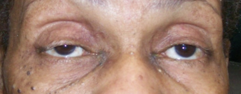
Multiple pulmonary pathologies—such as lung cancer in this patient’s case—can cause Horner’s syndrome.
Multiple pulmonary pathologies—such as lung cancer in this patient’s case—can cause Horner’s syndrome. Click image to enlarge.

Beyond the Cookbook

All optometrists can recite the classic triad of –osis findings associated with Horner’s syndrome: ptosis, pupillary miosis, facial anhidrosis. Some cases can also include heterochromia of the iridies if the syndrome developed prior to two years of age.1-8 Often, patients will display a dilation lag in dim illumination; that is, the affected pupil will slowly dilate and the anisocoria will diminish after several minutes in a dark room.
However, not every case of Horner’s syndrome will manifest classically; a “cookbook” approach to diagnosis is insufficient. It is incumbent on the clinician to use every clue in the presentation to suspect Horner’s syndrome may be present. In such cases, the dilation lag may be quite helpful in prompting pharmacologic testing.
Knowledge of the relevant anatomy is critical to localize the cause of confirmed Horner’s syndrome. 
A three-neuron arc provides sympathetic innervation to the eye.1-8 Horner’s syndrome is an interruption along this oculosympathetic pathway, between its origin in the hypothalamus and the ipsilateral dilator muscle of the eye.1-8 The first-order neuron originates in the hypothalamus, where it descends through the brain stem to the ciliospinal center of Budge, between C8 and T4 of the spinal cord.1-9 At this level, it synapses with the second-order neuron whose preganglionic cell bodies give rise to fibers that exit the spinal cord through the ventral spinal root; from here, they pass over the apex of the lung and enter the sympathetic chain in the neck, where they synapse in the superior cervical ganglion.1-8 
There, in the superior cervical ganglion, cell bodies of third-order neurons give rise to postganglionic fibers that travel with the internal carotid artery through the cavernous sinus towards the eye.1-9 These postganglionic fibers form the long posterior ciliary nerves, which course with the branches of the fifth cranial nerve to innervate the iris dilator muscle, Müller’s muscle and lacrimal gland.1-9 
Additionally, postganglionic sympathetic fibers, responsible for facial sweating, follow the external carotid artery to the sweat glands of the face.1-8 

Pharmacological Testing

Historically, topical liquid 10% cocaine was used to identify the presence of Horner’s syndrome, and topical hydroxyamphetamine was used to aid in localizing a postganglionic lesion.4-8,10 Today, these agents are not practically available; instead, the diagnosis of a suspected Horner’s syndrome can be confirmed by pharmacological testing with apraclonidine 0.5% or 1%.4-8,10 
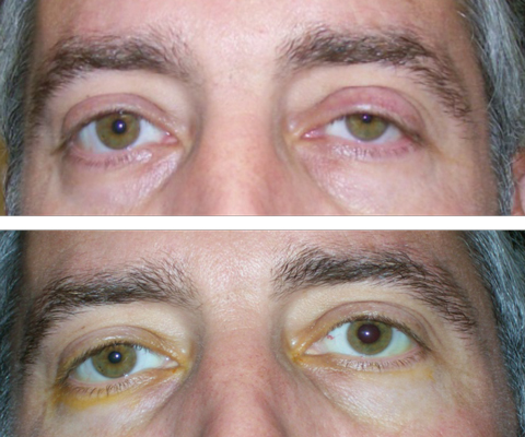
In Horner’s syndrome, the affected pupil and levator undergo denervation hypersensitivity.10-15 Apraclonidine is an alpha-2 adrenergic agonist that also acts as a very weak alpha 1-adrenergic agonist; when topically applied in Horner’s syndrome, it causes dilation of the affected pupil and possibly lid elevation, without change to the normal pupil.10-15 These changes are so dramatic that a ‘reversal’ occurs; the normal pupil and lid now appear ptotic and miotic in comparison with the apraclonidine-positive eye. 
Although sensitivity of apraclonidine testing is very good, false negative testing with apraclonidine may occur in acute Horner’s syndrome as the test relies on denervation hypersensitivity, which may not have had time to occur.12,13However, there are reports of positive apraclonidine tests within a few hours of the onset of symptoms related to Horner’s syndrome.14,15 Again, if the findings are equivocal but suspicion still high, one must go with clinical intuition and investigate further.
At top, this patient presented with suspected left Horner’s syndrome, pre-apraclonidine testing. At bottom, post-apraclonidine testing led to reversal, confirming the diagnosis.
At top, this patient presented with suspected left Horner’s syndrome, pre-apraclonidine testing. At bottom, post-apraclonidine testing led to reversal, confirming the diagnosis.Click image to enlarge.

Finding the Cause

Apraclonidine testing can easily confirm the diagnosis of Horner’s syndrome, but is unable to localize the lesion. Due to the long course of the oculosympathetic pathway from hypothalamus to the eye, there are multiple locations of potential pathology in Horner’s syndrome.16 Common etiologies include, but are not limited to: carotid artery dissection, aortic dissection, trauma, demyelinating disease such as multiple sclerosis (MS), tuberculosis, sarcoidosis, cluster headache, Pancoast syndrome, herpes zoster, giant cell arteritis (GCA) and malignancy.1-8,16,17 A careful physical exam and patient history can help guide imaging studies to aid in determining the location of the lesion.16
Once Horner’s syndrome is diagnosed (such as with a positive apraclonidine test), a directed evaluation is always preferable to non-specified testing. This is where knowledge of anatomy, symptoms and associated finding becomes crucial. For example, if there is a positive history of neck or facial pain, headache, recent neck trauma, ipsilateral vision loss or transient numbness or weakness on the opposite side of the body, an asutute clinician should suspect acute carotid artery dissection and order emergent neuroimaging.18-22 
In cases caused by internal carotid artery dissection, significant risk exists for major stroke within the first two weeks of onset.18-20 Computed tomography (CTA) or magnetic resonance angiography (MRA) of the neck and cervical spine, which must include Circle of Willis and the orbits down to the level of the aortic arch (T4-T5), which includes the carotid arteries and intracranial vessels as well as lung apices, is recommended.16,22 For cervical arterial dissections, CTA is the preferred imaging modality as, though MRI shows resolution with high contrast, it has lower temporal and spatial resolution.23 
In cases involving acute pain with onset of Horner’s syndrome, the patient requires emergency referral with imaging performed on the same day as clinical evaluation.16 Preferably, practitioners should not try to obtain this imaging themselves; rather, they should send the patient directly to the emergency room with detailed notes on the diagnosis of Horner’s syndrome and the clinical suspicion of carotid dissection. If they have just performed apraclonidine testing on the patient and they note apparent pharmacologic ‘reversal’ of the Horner’s syndrome, they must explain everything clearly in the referral notes so that the emergency room physicians focus on the correct anatomical side.
Carotid artery dissection, which affects the third-order neuron, can result from relatively minor trauma, or can occur spontaneously in patients with histories of connective tissue disease such as Ehlers-Danlos syndrome, Marfan’s syndrome or those with long-standing hypertension.1-8,24
In patients with histories of tobacco use, ipsilateral shoulder or arm pain or muscle weakness of the hand and arm, localization could lead to a lesion at the apex of the lung.25,26 As the oculosympathetic plexus courses over the apex of the lung via the second-order neuron, multiple pulmonary pathologies can cause Horner’s syndrome. For instance, Pancoast syndrome is a malignancy of the superior pulmonary sulcus most often caused by non-small cell lung carcinoma.25 Pancoast tumor has a rapid and high mortality rate.25 
Neuroradiological evaluation, typically including MRI of the chest and neck with and without contrast, and physical examination, often performed in concert with an internist, may lead to other chest and lung pathologies, including sarcoidosis or tuberculosis.16,26 Suspicion of cancer warrants a referral to primary care.

After Apraclonidine: The Next Steps

Once the apraclonidine test is positive for Horner’s syndrome, here are the next four steps in your clinical workup:
Step 1: Probe the patient’s history to see if the lesion can be localized. Any head, neck or eye pain can be suspicious for internal carotid artery dissection. Facial weakness or numbness generally localize to abnormalities within the middle cranial fossa. Tobacco history can cause concern for cancer at the lung apex. Other lung pathology may indicate sarcoidosis or tuberculosis. Dermatologic vesicles and neuralgia can identify zoster as a cause. Cluster migraines also can cause Horner’s syndrome. Thus, a physical examination and history, often done in concert with an internist, help dictate management.
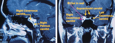
Sagittal MRI (left) shows an Internal carotid artery dissection in the wall of the petrous and cavernous sinus segments. Coronal MRI (right) shows significant narrowing of the carotid lumen in the cavernous sinus and supraclinoid segments of the right Internal carotid artery.
Sagittal MRI (left) shows an Internal carotid artery dissection in the wall of the petrous and cavernous sinus segments. Coronal MRI (right) shows significant narrowing of the carotid lumen in the cavernous sinus and supraclinoid segments of the right Internal carotid artery. Click image to enlarge.
Step 2: Look for other neurologic signs. Cranial nerve VI palsy in association with Horner’s identifies cavernous sinus as area of interest, but does not rule out a brain stem lesion. Current or past history of optic neuritis can indicate MS.
Step 3: Look at the age of the patient. Young people can suffer from MS and older people can suffer from stroke. Always assess GCA risk in patients older than 60.
Step 4: Obtain the appropriate imaging. It’s best to image the area of suspicion as identified above. If that is unrevealing, the patient must be imaged from chest to head including MRA or CTA of neck for carotid dissection, MRI of brain with and without contrast with attention to middle cranial fossa and cavernous sinus, and chest/neck MRI or CT to include the brachial plexus and cervical spine. Non-targeted evaluations are often non-productive, but still recommended to rule out treatable pathology. Unless the patient has an emergency situation regarding carotid dissection, consider referring to a primary care physician or neurologist for an overall health evaluation with consideration for these conditions.

Other Neurological Signs

In patients presenting with abducens (cranial nerve VI) palsy in addition to Horner’s syndrome, clinicians should pay attention to the cavernous sinus.5,27The oculosympathetic plexus travels with the abducens nerve within the cavernous sinus.5,27,28 MRI of the brain with and without contrast, with attention to the middle cranial fossa and cavernous sinus, is warranted. 
However, lesions of the first-order neuron including brain stem lesions in Horner’s syndrome may also cause abducens nerve palsy, so additional clinical signs and symptoms can aid in localization. Patients with first-order neuron Horner’s syndrome typically present with widespread neurological symptoms including contralateral hemiparesis, contralateral hypesthesia and hypohidrosis of one side of the body.16,29 Pontine lesions such as stroke may additionally result in dizziness, nausea and difficulty swallowing.29,30 In cases of nystagmus, vertigo and vomiting—in addition to Horner’s syndrome, lateral medullary infarction or Wallenburg syndrome—may be present.29 Horner’s syndrome with trochlear (cranial nerve IV) palsy suggests the dorsal mesencephalon is an area of interest for clinicians to investigate.29
In cases of Horner’s syndrome, consider the age of the patient. In younger patients, demyelinating disease, including MS, can be a potential cause of Horner’s syndrome. In older patients, stroke, zoster and GCA are etiologies that practitioners should consider.21,32
When no diagnostic clues are identified following a complete clinical examination, a non-targeted evaluation, including imaging of the upper chest, neck, and brain, must be performed.16,17,22 MRI of the neck and chest including the lung apex, MRA or CTA of the neck and cervical spine, and MRI of the brain, with attention to the middle cranial fossa, is recommended. Imaging centers may have a protocol for dealing with Horner’s syndrome to ensure that proper studies are done. In many cases, even with extensive imaging, an underlying cause may not be determined.9,16,17     
With any radiology referral, it is important to provide the neuroradiologist with all available localizing information available—including the  anatomical location of Horner’s syndrome—to allow for clinical-radiologic correlation.11 Remember, a neuroradiologist cannot help you if you do not:
  1. order a scan,
  2. order the correct scan, and 
  3. specify what you are looking for.
Once Horner’s syndrome has been identified with a positive apraclonidine test, the real work begins. Optometrists must use their knowledge of anatomy and rely on the history and clinical exam to determine potential causes, send to an emergency room when appropriate, order necessary diagnostic testing and comanage with other medical specialists when necessary.
Dr. Steen is an attending optometrist and assistant professor of ocular pharmacology at Nova Southeastern University’s College of Optometry. 
Dr. Sowka is a professor at Nova Southeastern University.
View Footnotes